Nov 21, 2012

Tooth Extraction

Patient:  X.V
Age: 50
Diagnostic:
     - Partial edentulism 
     - Periodontal disease  
     - Calculum supragingival and subgingival

Our patient has:

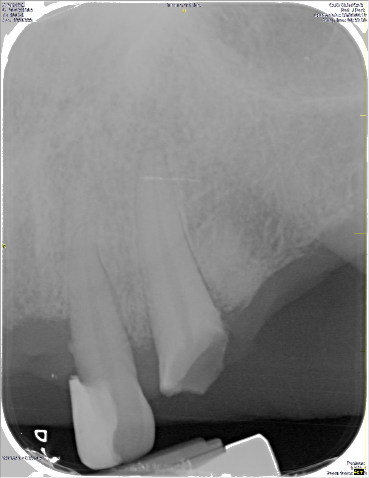
   - Superior arch: second right molar, both canines and lateral incisors all with big restorations. Plus a partial superior prosthesis supported by the fractured canine and the molar.
   - Lower arch: the patient has in relative good condition from the first right premolar to the second left premolar and no prosthesis.   


Treatment plan:
- First phase: hygienic phase. We proceeded to do a tartrectomy.
- Second phase: Surgical phase. We removed the superior left canine (2.3) that had a crown fracture. 
- Third phase: prosthodontic phase. Reemplace missing tooth. For now the patient don't want to do the lower prosthesis because she can't afford it. We are waiting if one day is ready. 


Sep 5, 2012

4th year

After a long long summer is time to get ready into my 4th year at college. This is my new schedule for this year. What a mess!!! 


May 16, 2012

Closed for exams!

I know it's been a long time but exams are coming. Ill start next Monday 21rst with periodontics, then 23rd with paediatric dentistry and for the 25th Oral medicine. Then last week with clinic introduction, prosthodontics and orthodontics. 


This is just Oral medicine (Surgery):


Wish me good luck!!!

Apr 17, 2012

Set up

The set up is the best test for the diagnosis. It consists in divide and relocation the teeth of the models. 

There is a lot of ways to do it like for example by computer programs in 3D or the manual way. Here in school they prefer to do with our hands, it takes for 4 hours more or less. 
First of all we need to plan witch teeth we are going to remove for a good occlusion Class I. We want to remove all the first premolar so we can have space for all the teeth. 


We need to make two holes. The first one under the second premolar and first molar. The second hole is under the two central incisors. 


We introduce hacksaw between the holes. 


Finally we are going to have 5 teeth in total for the first part. We have to divide teeth by teeth without destroying the contact point. Then we start mounting the teeth with wax without the first premolar. That's very important.  


And like this, we continue. 


Mar 29, 2012

Magnetic resonance imaging of TMJ

Magnetic resonance imaging (MRI) of my Temporomandibular joint.

After to remove my wisdom teeth I started having jaw pain and articular problems. Now, 2 years later more or less, I did a MRI. Is a diagnostic prove that shows soft tissues. 

Left side: Seems good, just a small dislocation. 


Right side: displacement of the disc


If we take a look at the right side: the red circle shows the disc. We can see a displacement of the disc without reuptake. If it was good the disc will be in the glenoid fossa. 


In the coronal section we can see that if we go forward, the disc is going to be before the fossa not with the condile. 


Mar 20, 2012

CASE 4563.2

Yesterday at the university clinic, a patient came for a cleaning. He said he had no apparently disease and that he also brushes his teeth once a day.
He has a severe gingivitis generalised. In the first visit this patient was bleeding all the time so the probing was very difficult. He has a lot of calculus under his gums that apparently we can not see only deduce because his gums are red, shining and big.
We tried to discover why, brushing his teeth once a day, he had this gingivitis. In one moment he said that he spent 2 years in prison and that's why he couldn't brush his teeth well. He lived under pressure.  

Here is the case before we started: 


Here is the end:


Now we need to visit him next time to see if his gums disminued with the cleaning and the products that we gave to him: 


Mar 18, 2012

Pediatric Crown

Pediatric Crown are the best durable restoration and successful in primary dentition with decays or fractures. It doesn't break and rarely wears in the few years remaining in the mouth. It remains attached to the tooth until the exfoliation and you always have to inform the parents about this fact.

It is indicated in this cases:
  • Restoration of cavities in two or more surfaces
  • Children with high risk of cavities
  • After receiving a pulp treatment: pulpotomy or pulpectomy. 
  • Teeth with structural defects
  • Restoration in fractured teeth or fractured cusps
  • Used like a space maintainer and teeth with excessive wear.
In this image we can see the crowns with the mainteiner.
 

It is contraindicated when the teeth is going to be exfoliated in less than 6-12 months. 

They provide us with this metallic crown:


We first have to isolate the teeth and then we start milling: